Teaching Note 1 of 5
No Attempts Yet
Awaiting Selection
Review Malleus handle
Select pin 1 in the image, then complete the activity.
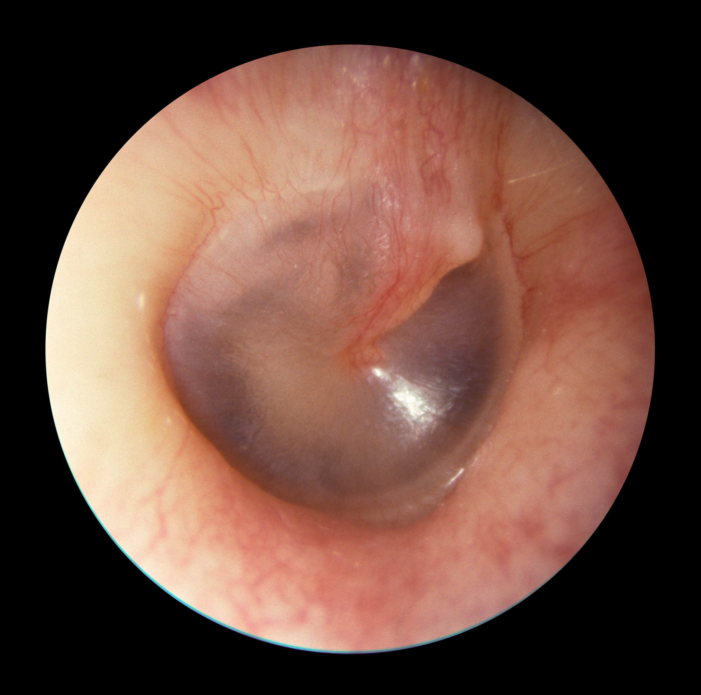
The handle of the malleus is a key landmark that helps orient the tympanic membrane.
Use the marker in the image to open its activity.
Use the media surface and panel to continue.
Target
- Focus the target, review the note, then mark it reviewed.
Selected
Review This Teaching Point, Then Mark Reviewed.
Review this teaching point.
Mobile tip: media stays in focus. Expand the task sheet for answers, hints, and review.
The handle of the malleus is a key landmark that helps orient the tympanic membrane.
Teaching Note 2 of 5
No Attempts Yet
Awaiting Selection
Review Umbo
Select pin 2 in the image, then complete the activity.
The umbo is the central point where the malleus attaches to the tympanic membrane.
Use the marker in the image to open its activity.
Use the media surface and panel to continue.
Target
- Focus the target, review the note, then mark it reviewed.
Selected
Review This Teaching Point, Then Mark Reviewed.
Review this teaching point.
Mobile tip: media stays in focus. Expand the task sheet for answers, hints, and review.
The umbo is the central point where the malleus attaches to the tympanic membrane.
Teaching Note 3 of 5
No Attempts Yet
Awaiting Selection
Review Cone of light
Select pin 3 in the image, then complete the activity.
The cone of light is usually seen in the anteroinferior quadrant of a normal tympanic membrane.
Use the marker in the image to open its activity.
Use the media surface and panel to continue.
Target
- Focus the target, review the note, then mark it reviewed.
Selected
Review This Teaching Point, Then Mark Reviewed.
Review this teaching point.
Mobile tip: media stays in focus. Expand the task sheet for answers, hints, and review.
The cone of light is usually seen in the anteroinferior quadrant of a normal tympanic membrane.
Teaching Note 4 of 5
No Attempts Yet
Awaiting Selection
Review Pars tensa
Select pin 4 in the image, then complete the activity.
The pars tensa forms the larger, tense portion of the tympanic membrane.
Use the marker in the image to open its activity.
Use the media surface and panel to continue.
Target
- Focus the target, review the note, then mark it reviewed.
Selected
Review This Teaching Point, Then Mark Reviewed.
Review this teaching point.
Mobile tip: media stays in focus. Expand the task sheet for answers, hints, and review.
The pars tensa forms the larger, tense portion of the tympanic membrane.
Teaching Note 5 of 5
No Attempts Yet
Awaiting Selection
Review Pars flaccida
Select pin 5 in the image, then complete the activity.
The pars flaccida is the smaller superior portion and is clinically important when assessing retraction pockets.
Use the marker in the image to open its activity.
Use the media surface and panel to continue.
Target
- Focus the target, review the note, then mark it reviewed.
Selected
Review This Teaching Point, Then Mark Reviewed.
Review this teaching point.
Mobile tip: media stays in focus. Expand the task sheet for answers, hints, and review.
The pars flaccida is the smaller superior portion and is clinically important when assessing retraction pockets.